Hi-Fi (f)MRI: a high-resolution, fast-sampling MRI framework in humans for eye and brain imaging at 3T
Hi-Fi (f)MRI: a high-resolution, fast-sampling MRI framework in humans for eye and brain imaging at 3T
Prof. Dr. Benedetta Franceschiello
Translational Imaging Center (TIC)
sitem-insel Bern
Wednesday, 04 December 2024, 16:00
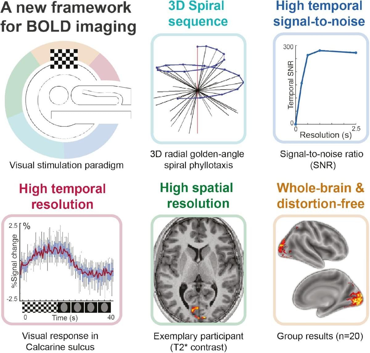
In the fields of medical diagnostics and neuroscience, Magnetic Resonance Imaging (MRI) and functional magnetic resonance imaging (fMRI) are foundational technologies. MRI, a powerful and non-invasive instrument, plays a pivotal role in medical practice by revealing tissue properties and diagnosing pathologies. However, when applied to dynamic organs like the eyes, MRI faces challenges primarily driven by eye-motion artifacts, which constitute the primary limitation to the use of MRI in ophthalmology. To address these challenges and inspired by recent advancements in the field of cardiac MRI, we have introduced a Motion- Resolved 3D MRI technology for eye-imaging, providing a pioneering solution that eliminates the need for patient immobilization and anesthesia. This innovation revolutionizes the applicability of MRI in ophthalmology, particularly in cases where oculomotricity is impaired, such as in strabismus. Motion-related issues also impact the usability of fMRI in neuroscience research. fMRI takes a central role in the study of the brain, particularly in investigating cognitive functions. Traditional methods using echo-planar imaging (EPI) involve trade-offs between spatial and temporal resolutions. In contrast, we will discuss how the same approach used to address motion-related challenges in eye imaging can be extended to the brain, overcoming these limitations. This novel methodology offers higher spatial resolution (1mm³) and improved temporal precision (up to 250ms) with comprehensive whole-brain coverage. It corrects artifacts before image reconstruction and allows post-scan adjustments of temporal resolution without presuming a specific hemodynamic response shape. Its reliability is confirmed through an experiment involving 20 participants performing an ON-OFF visual paradigm, demonstrating its effectiveness in cognitive neuroscience research.
The lecture will be held as in-person meeting at sitem-insel (Room O2.211), Freiburgstr. 3, Bern (followed by an Apéro to continue discussions) and in addition broadcast via Zoom@
and do spread the word to anybody potentially interested (for further info: bernd.jung@insel.ch).