Our Partners
The Translational Imaging Center (TIC) is part of the Inselgruppe AG, the largest and leading medical care system in Switzerland, specifically as a unit within the Department Teaching and Research of the University Hospital. The realization of such a platform is only possible with the support of our key partner organizations, namely the Swiss Institute for Translational and Entrepreneurial Medicine (sitem-insel AG), a National Center of Excellence that assists in the transition from research findings or prototypes to marketable products, the University of Bern with its 150 institutes and nine inter- and transdisciplinary competence centers and Siemens Healthineers, a world-leading medical device company, that specialises in medical imaging equipment and diagnostics.


Organisation

Management

Research

Boards
Research
Shaping the future of MR imaging research in Switzerland
MRI technology continues to develop rapidly, and increasingly specialized applications have opened up an environment for comprehensive synergistic research areas consisting of life science, medicine and other basic research, including magnetic resonance methods, system biology and biomedical engineering. Working groups from the University Hospital Bern (Inselspital) and the University of Bern have joined forces to form an MRI research consortium with the goal to develop an integrative platform for translational research and education. The platform constitutes one of the core enabling facilities at sitem-insel and aims at leveraging the research from the cellular level up to clinical research. Close collaboration with our industry partners enables the development of novel MRI methods and applications and their translation into advanced and personalized patient care.

Imaging
Imaging Core Facilities

Basic Science
Basic Science & University

Neuroscience
Life & Neurosciences

Medicine
Biomedicine, Physiology & Surgery
Services
With its many collaboration partners of our MR consortium the TIC provides services and consulting for basic research, clinical research and epidemiological studies with emphasis on imaging. Research encompasses life science, psychiatric research, neurovascular and cardiovascular research, neuro-oncology, pre-surgical epilepsy research, elastography of the liver and many more. Services include consultancy with respect to imaging protocols, sequence optimization, imaging processing and data analysis. Other services can be provided through close cooperation with the clinical trial unit of the Faculty of Medicine of the University of Bern and the Bern University Hospital, Inselspital.

Project Planning

Study Hosting

Study Follow-Up
Events
The Translational Imaging Center hosts scientific and translation seminars and offers Continued Professional Development training events.

7T Balanced Steady State Free Precession:
Theory and Applications from Low to High-Field
20 March 2024

New technology in designing human head RF coils for UHF MRI: Dipole antennas
24 January 2024

fMRI at UHF: Perspectives for basic and clinical neuroscience
15 November 2023
News
Updates and Achievements

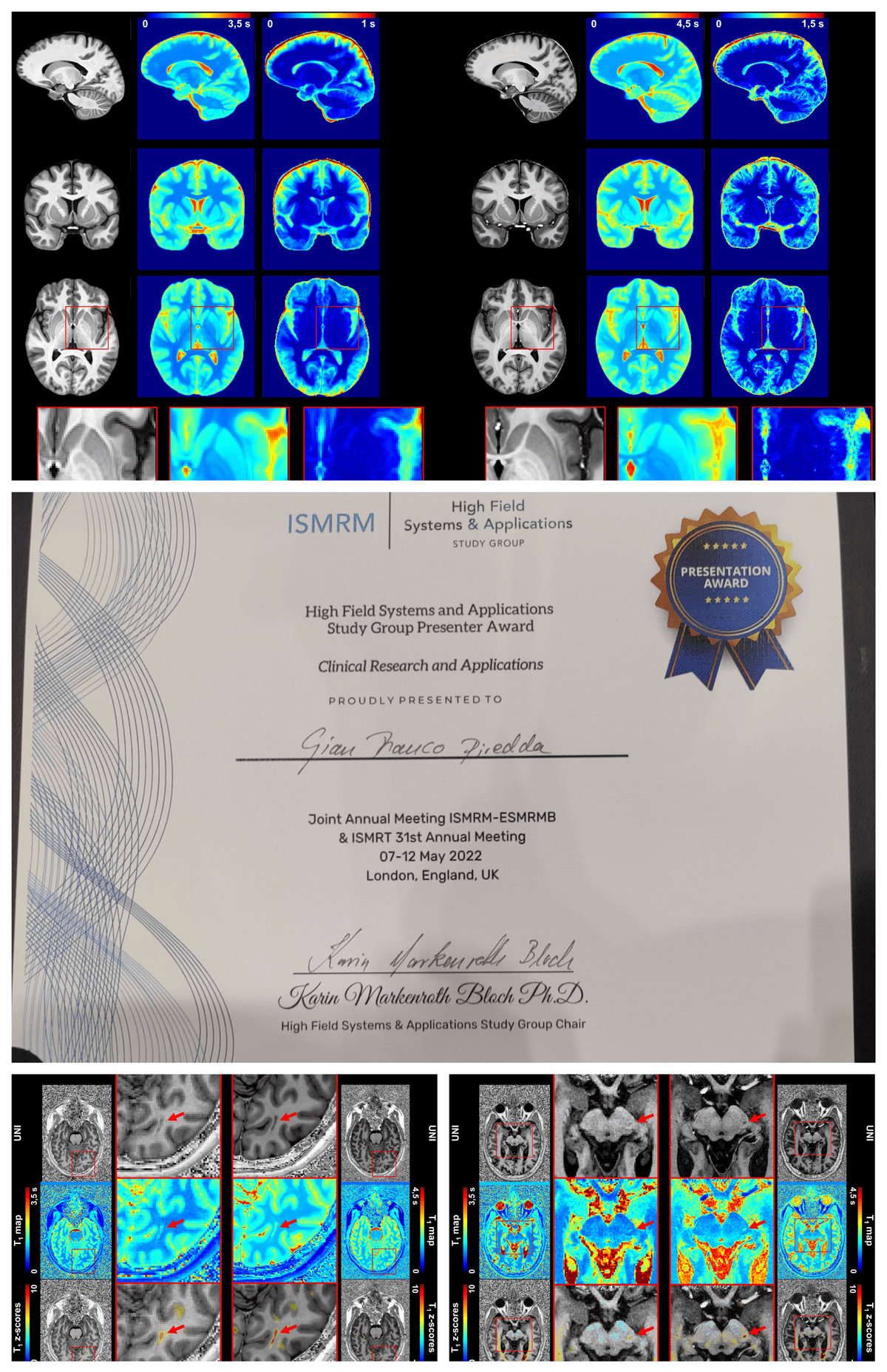
Magnetic Resonance in Medicine: April 2023 Editor's Pick
"Submillimeter T1 atlas for subject-specific abnormality detection at 7T"
Magnetic Resonance in Medicine April 2023 Editor's PickG. Piredda1,2,3, S. Caneschi1,4,5, T. Hilbert1,4,5, G. Bonanno6,7,8, A. Joseph6,7,8, K. Egger9, J. Peter10, S. Klöppel10, E. Jehli11,12, M. Grieder11, J. Slotboom13, D. Seiffge14, M. Goeldlin14, R. Hoepner14, T. Willems15, S. Vulliemoz16, M. Seeck16, P.B. Venkategowda17, R.A. Corredor Jerez1,4,5, B. Maréchal1,4,5, J.-P. Thiran4,5, R. Wiest7,13, T. Kober1,4,5, P. Radojewski7,13
Purpose
Studies at 3T have shown that T1 relaxometry enables characterization of brain tissues at the single-subject level by comparing individual physical properties to a normative atlas. In this work, an atlas of normative T1 values at 7T is introduced with 0.6 mm isotropic resolution and its clinical potential is explored in comparison to 3T.
Methods
T1 maps were acquired in two separate healthy cohorts scanned at 3T and 7T. Using transfer learning, a template-based brain segmentation algorithm was adapted to ultra-high field imaging data. After segmenting brain tissues, volumes were normalized into a common space, and an atlas of normative T1 values was established by modeling the T1 inter-subject variability. A method for single-subject comparisons restricted to white matter and subcortical structures was developed by computing Z-scores. The comparison was applied to eight patients scanned at both field strengths for proof of concept.
Results
The proposed method for morphometry delivered segmentation masks without statistically significant differences from those derived with the original pipeline at 3T and achieved accurate segmentation at 7T. The established normative atlas allowed characterizing tissue alterations in single-subject comparisons at 7T, and showed greater anatomical details compared with 3T results.
Conclusion
A high-resolution quantitative atlas with an adapted pipeline was introduced and validated. Several case studies on different clinical conditions showed the feasibility, potential and limitations of high-resolution single-subject comparisons based on quantitative MRI atlases. This method in conjunction with 7T higher resolution broadens the range of potential applications of quantitative MRI in clinical practice.
1Advanced Clinical Imaging Technology, Siemens HealthineersInternational AG, Lausanne, Switzerland
2Human Neuroscience Platform, Fondation Campus Biotech Geneva,Geneva, Switzerland
3CIBM-AIT, École Polytechnique Fédérale de Lausanne (EPFL),Lausanne, Switzerland
4Department of Radiology, Lausanne University Hospital andUniversity of Lausanne, Lausanne, Switzerland
5LTS5, École Polytechnique Fédérale de Lausanne (EPFL), Lausanne,Switzerland
6Advanced Clinical Imaging Technology, Siemens HealthineersInternational AG, Bern, Switzerland
7Translational Imaging Center (TIC), Swiss Institute for Translationaland Entrepreneurial Medicine, Bern, Switzerland
8Magnetic Resonance Methodology, Institute of Diagnostic andInterventional Neuroradiology, University of Bern, Bern, Switzerland
9Department of Neuroradiology, Faculty of Medicine, University ofFreiburg, Freiburg, Germany
10University Hospital of Old Age Psychiatry and Psychotherapy,University of Bern, Bern, Switzerland
11Translational Research Center, University Hospital of Psychiatry andPsychotherapy, University of Bern, Bern, Switzerland
12Department of Neurosurgery, University Hospital of Zurich, Zurich,Switzerland
13Support Center for Advanced Neuroimaging, Institute for Diagnosticand Interventional Neuroradiology, Inselspital, University of Bern,Bern, Switzerland
14Department of Neurology, University Hospital Bern, Inselspital,University of Bern, Bern, Switzerland
15Institute of Psychology, University of Bern, Bern, Switzerland
16EEG and Epilepsy Unit, Department of Clinical Neurosciences, GenevaUniversity Hospitals and Faculty of Medicine, Geneva, Switzerland
17Siemens Healthcare Pvt. Ltd., Bangalore, India
Access free here https://onlinelibrary.wiley.com/doi/10.1002/mrm.29540

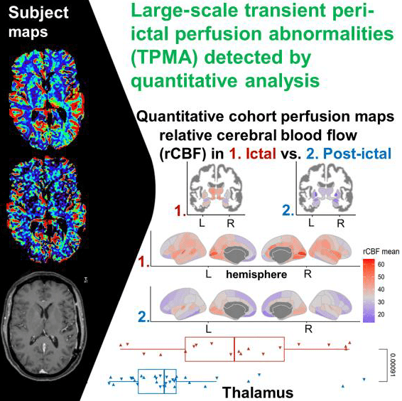
Detecting imaging abnormalities in epilepsy using quantitative image analysis
"Large-scale transient peri-ictal perfusion magnetic resonance imaging abnormalities detected by quantitative image analysis"
M. Köstner1,2, M. Rebsamen1,3, P. Radojewski1,4, C. Rummel1, B. Jin5, R. Meier1, U. Ahmadli1, K. Schindler5, and R. Wiest1,4
Brain Communications February 2023Epileptic seizures require a rapid and safe diagnosis to minimize the time from onset to adequate treatment. Some epileptic seizures can be diagnosed clinically with the respective expertise. For more subtle seizures, imaging is mandatory to rule out treatable structural lesions and potentially life-threatening conditions. MRI perfusion abnormalities associated with epileptic seizures have been reported in CT and MRI studies. However, the interpretation of transient peri-ictal MRI abnormalities is routinely based on qualitative visual analysis and therefore reader dependent. In this retrospective study, we investigated the diagnostic yield of visual analysis of perfusion MRI during ictal and postictal states based on comparative expert ratings in 51 patients. We further propose an automated semi-quantitative method for perfusion analysis to determine perfusion abnormalities observed during ictal and postictal MRI using dynamic susceptibility contrast MRI, which we validated on a subcohort of 27 patients. Visual ratings between expert readers performed well on the patient level, but visual rating agreement was low for analysis of subregions of the brain. The asymmetry of the automated image analysis correlated significantly with the visual consensus ratings of both readers. We conclude that expert analysis of dynamic susceptibility contrast MRI effectively discriminates ictal versus postictal perfusion patterns. Automated perfusion evaluation revealed favourable interpretability and correlated well with the classification of the visual ratings. It may therefore be employed for high-throughput, large-scale perfusion analysis in extended cohorts, especially for research questions with limited expert rater capacity.
1Support Center for Advanced Neuroimaging (SCAN), University Institute of Diagnostic and Interventional Neuroradiology,
University of Bern, Inselspital, Bern University Hospital, Bern CH-3010, Switzerland
2Faculty of Medicine, University of Bern, Bern CH-3008, Switzerland
3Graduate School for Cellular and Biomedical Sciences, University of Bern, Bern CH-3012, Switzerland
4Translational Imaging Center (TIC), sitem-Insel, Bern University Hospital, Bern CH-3010, Switzerland
5Department of Neurology, Inselspital, Sleep-Wake-Epilepsy-Center, Bern University Hospital, University of Bern, Bern CH-3010, Switzerland
Access free here https://academic.oup.com/braincomms/article/5/2/fcad047/7055970

Recognition of TIC Research at ISMRM &ISMRT 2023 Toronto
The TIC team is thrilled to announce that the Translational Imaging Center has achieved a significant international success, with a total of 31 scientific peer-reviewed abstracts accepted to be presented at the International Society for Magnetic Resonance in Medicine, 03 - 08 June 2023, Toronto. The research reported covers MRI developments across many organ systems from head to toe, and magnetic field strengths from 1.5T to 7T. The topics of the abstracts range from diffusion-weighted MR spectroscopy of the prostate to the detection of flow in the heart and neuronal currents. The presentations cover 10 oral contributions, 4 power pitches, and numerous posters. The success at this leading scientific congress in the field of MRI is a testament to the TIC's dedication and expertise in the field of noninvasive medical imaging. It demonstrates the TIC's commitment to continuing to push the boundaries of what is possible with MRI technology.
Link to ISMRM website: 2023 ISMRM Annual Meeting & Exhibition – ISMRM

Better imaging of Multiple Sclerosis patients
"Reliable brain morphometry from contrast-enhanced T1w-MRI in patients with multiple sclerosis"
M. Rebsamen, R. McKinley, P. Radojewski, M. Pistor, C. Friedli, R. Hoepner, A. Salmen, A. Chan, M. Reyes,
F. Wagner, R. Wiest, C. Rummel
Human Brain Mapping October 2022Brain morphometry is usually based on non-enhanced (pre-contrast) T1-weighted MRI. However, such dedicated protocols are sometimes missing in clinical examinations. Instead, an image with a contrast agent is often available. The methodology described demonstrates that brain morphometry can be derived reliably from contrast-enhanced MRI using Deep-Learning-based morphometry tools, making additional cases available for analysis and potential future diagnostic morphometry tools.
Access free here https://doi.org/10.1002/HBM.26117

Prof. Dr. med. Roland Wiest speaks at INGE St. Symposium 2022
"New possibilities for neuroscience with 7 Tesla MRI"
The INGE St. 2022 symposium explored "New possibilities for neuroscience through 7 Tesla MRI".
Use of Ultra-high-field MRI in the most recent clinical and neuroscientific applications will be highlighted by internationally renowned speakers to foster discussions on how 7 Tesla MRI can advance neuroscience research. The programme harnesses the interaction of researchers from a variety of fields including medicine, cognitive sciences and MR physics to benefit all interested communities.
More information here INGE St. Symposium 2022

Prof. Dr. sc. nat. Roland Kreis awarded with the honorary membership of ESMRMB
This was announced at the recent joint congress of ISMRM and ESMRMB in London. Honorary membership is the highest award granted by the European Society for Magnetic Resonance in Medicine and Biology, the largest society for MR research in Europe. The award was bestowed upon Prof Kreis from the MR Methods Group for his outstanding contributions to research in clinical magnetic resonance, in particular quantitative MR spectroscopy, but also for his longstanding commitment to the society.

Prof. Dr. rer. soc. Katharine Henke receives SNSF Advanced Grant
"Strengthening memory formation for retrieval despite amnesia"
Prof. Dr. rer. soc. Katharine Henke, Professor of Experimental Psychology and Neuropsychology at the Institute of Psychology, University of Bern and Delegate of the Psychology group of the Neuroscience Cluster of the TIC sitem has been awarded SNSF Advanced Grant funding of CHF 1.5 Mio over 5 years.
Due to the demographic change, the prevalence of amnesia associated with old age rises. Amnesia is a deficit of episodic memory, which is believed to result from a loss of episodic learning. Yet, research in mice suggests that new episodic memories continue to be formed in amnesia because physical memory traces of newly formed memories were discovered. These memories harboured the complete information content, although they were not retrievable. The physical memory traces were reactivated optogenetically to the point where the memories became consciously retrievable despite amnesia. Hence, learning had survived in residual intact hippocampal-neocortical tissue besides pathology in amnesic mice. No one has tracked conscious and unconscious episodic memories over time in healthy and amnesic humans because the necessary techniques were missing and because traditional memory theories claim that episodic memories are bound to be consciously accessible or are inexistent. Tracking newly formed individual episodic memory traces with high-resolution functional magnetic resonance imaging in healthy individuals and amnesic patients is now feasible in Bern with the advent of ultra high-field magnetic resonance imaging. I will use ultra high-field functional magnetic resonance imaging to track memory traces underlying individual episodic memories in healthy and amnesic humans. We will track the dynamic transitions of newly formed memories and their underlying physical memory traces as these memories change their representational status und the geometric configuration of their traces over the 24 hours following episodic learning. During this consolidation time, some memories are expected to shift from being consciously accessible to being only unconsciously accessible and to being inaccessible (forgotten). We will demonstrate that the access to stored episodic memories can vary dynamically during the memories’ lifetime in healthy and amnesic individuals. Using electrical brain-stimulation we will reinforce the physical memory traces of newly formed memories in healthy and amnesic individuals to bring unconscious memories to conscious access. To uncover the mechanism of this strengthening of memory traces, we will use ultra high-field magnetic resonance spectroscopy. We expect brain stimulation-induced local glutamatergic increases to result from an enhanced long-term potentiation that results from electrical brain stimulation.

ISMRM Prize - Best abstract in "Clinical Research and Applications" at 7T
"High-resolution T1 atlas for subject-specific abnormality detection at 7T”
Gian Franco Piredda1,2,3, Piotr Radojewski4,5 ,Gabriele Bonanno5,6,7, Arun Joseph5,6,7, Karl Egger8, Shan Yang8, Punith B. Venkategowda9, Ricardo A. Corredor Jerez1,2,3, Bénédicte Maréchal1,2,3, Roland Wiest4,5, Jean-Philippe Thiran2,3, Tom Hilbert1,2,3, Tobias Kober1,2,3
1Advanced Clinical Imaging Technology, Siemens Healthcare AG, Lausanne, Switzerland,
2Department of Radiology, Lausanne University Hospital and University of Lausanne, Lausanne, Switzerland,
3LTS5, Ecole Polytechnique Federale de Lausanne (EPFL), Lausanne, Switzerland,
4Support Center for Advanced Neuroimaging, Institute for Diagnostic and Interventional Neuroradiology, Inselspital, Bern University, Bern, Switzerland,
5Translational Imaging Center (TIC), Swiss Institute for Translational and Entrepreneurial Medicine, Bern Switzerland
6Advanced Clinical Imaging Technology, Siemens Healthcare AG, Bern, Switzerland,
7Magnetic Resonance Methodology, Institute of Diagnostic and Interventional Neuroradiology, University of Bern, Bern, Switzerland,
8Department of Neuroradiology, Medical Center – University of Freiburg, Faculty of Medicine, University of Freiburg, Freiburg, Germany,
9Siemens Healthcare Pvt. Ltd., Bangalore, India.
A joint collaboration was awarded best abstract at the 2022 ISMRM Prize in the category of “Clinical Research and Applications” of the High Field Systems and Applications study group. MRI studies conducted at 3T have shown that T1 relaxometry enables personalized characterization of brain tissues by comparing physical properties of a single patient to a normative atlas. Ultra-high field imaging allows exploiting this concept at even higher resolutions, which can be crucial to detect certain diseases. In this study we established an atlas of normative T1 values at 7T from acquisitions with 0.6 mm isotropic resolution. We investigated the clinical potential of 7T vs. 3T imaging in case reports from patients scanned at both field strengths. Our findings suggest that quantitative MR imaging at 7T allows improving the characterization of tissue abnormalities on a single-subject basis compared to 3T.

Mission Deuterium
"Metabolic imaging of the brain at ultra high field MRI: feasibility study of spectroscopic methods for exploration of brain metabolism without ionizing radiation"
Principle Investigator: Prof. Johannes Slotboom of the SCAN/TIC, Neuroradiology
University Hospital Bern Switzerland
The aim of this Swiss National Science Foundation project is to study the metabolism in the aging brain. Glucose is the main source of energy if the human brain, and tight regulation of its metabolism is critical for brain physiology. Positron Emission Tomography (PET), which requires an injection of a radioactive substance, is the method most commonly used to study the uptake of glucose in the brain. Deuterium metabolic imaging (DMI) is a novel radiation-free magnetic resonance spectroscopic imaging approach to measure not only the glucose uptake in the brain, but also its metabolic products glutamate and lactate. Beyond glucose, important neurotransmitters (GABA and glutamate) will be studied using a novel approach based on proton magnetic resonance spectroscopic imaging recently developed by our group.
The research will be performed in collaboration of the Support Centre for Advanced Neuroimaging (SCAN), Institute for Diagnostic and Interventional Neuroradiology, Inselspital, Bern; Translational Imaging Center of the Sitem-insel AG; the Department of Diabetes, Endocrinology, Nutritional Medicine and Metabolism, Inselspital, Bern; University Hospital of Old Age Psychiatry and Psychotherapy, University of Bern, and the Department of Radiology, University of Miami School of Medicine, Miami, Florida, U.S.A. and the Radiology & Biomedical Imaging Group of the Yale School of Medicine, New Haven, U.S.A.

Spotting intracranial aneurysms with 7T MRI
"Clinical Implementation of 7T MRI for the Identification of Incidental Intracranial Aneurysms versus Anatomic Variants"
P. Radojewski, J. Slotboom, A. Joseph, R. Wiest and P. Mordasini
American Journal of Neuroradiology October 2021Most unruptured intracranial aneurysms can be adequately characterized using 1.5T and 3T MR imaging. Findings in a subgroup of patients can remain unclear due to difficulties in distinguishing aneurysms from vascular anatomic variants. We retrospectively analyzed clinical data from 30 patients with suspected incidental aneurysms on 3T MR imaging who underwent 7T MR imaging. Our findings suggest that 7T MR imaging provides a clarification tool for the group of patients with suspected unruptured intracranial aneurysms and diagnostic ambiguity after standard 3T MR imaging.
Access free here www.ajnr.org/content/42/12/2172